Iridology, Nutrition and Cognition Research

Current research in Iridology, Nutrition and Cognition of interest to the elderly and their family members, as well as to the Long Term Care industry as a whole.

Monday, February 20, 2006

The Classic 'Sub-acute Inflammation' Sign for an Under-acid Stomach
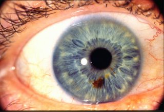
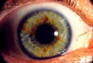
This is a picture of the classic 'sub-acute inflammation'--Bernard Jensen, 1952--iris sign indicative of an under-acid stomach: the light gray 'halo' which, in this picture, extends from 1-2 millimeters from the pupil.

Client had a very short attention span but was a genius at engineering, with several inventions over a variety of engineering disciplines. (Claimed that, if he had been 50 years younger, he would have been diagnosed with attention deficit hyperactivity disorder; which suggests that a calcium/magnesium deficiency may be an important aspect of such a diagnosis.)

Although having no knowledge or previous experience with medicine or physiology, client developed his own hydrochloric acid supplementation 'protocol', titrating the number of capsules based upon whether his sclera was either 'clear' or 'murky' in the morning. When his sclera was 'clear', he would take 3 2.5 grain capsules one or more times per day; when his sclera was 'murky' he would take as many as 8 2.5 grain capsules, once, twice or more times per day depending upon the effect upon his sclera. Had established a correlation between the condition of his sclera and his over-all feeling of well-being. ('Murkiness' to the sclera was likely a build-up of histamine; although this is merely conjecture.)

Within approximately a year of adopting this 'protocol', client reported that he had lost 22 pounds and was no longer taking prescription medications for either cholesterol, blood pressure or prostate problems.

This picture--specifically, the acute sign in the Peyer's Patches (2:30 o'clock, within the under-acid 'halo')--is, by the way, a graphic validation of an article entitled: "Local Hormone Networks and Intestinal T Cell Homeosotasis" published in SCIENCE, Vol. 275, 28 March,1997. (But an e-mail to the author in this regard failed to elicit a response.)
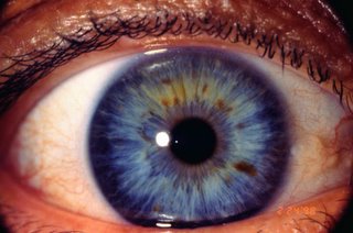
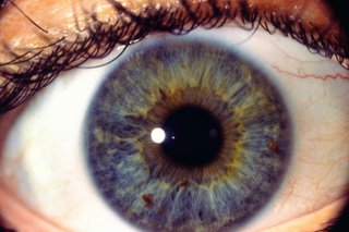
Acute Inflammation (Over-acid stomach) in a Blue Iris

The area of brightness immediately adjacent to the pupil either indicates a true over-acid stomach or is the result of dietary factors (jalapeno peppers, for example) and/or hypothyroidism. The yellow density--xanthelasmus--medial to the iris is suggestive of hyperlipidemia. (The client was an obese Hispanic woman.)

A Heidelberg test can be ordered by a doctor to test for stomach acidity. Also recommended: a lipid panel and tests for thyroid stimulating hormone (TSH), T4 and T3; and Reverse T3 to rule out Wilson's Thyroid Syndrome.

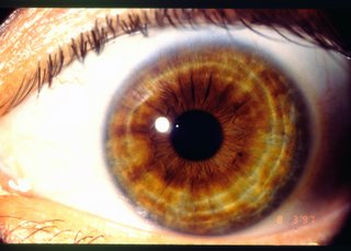
Sign for Over-acid Stomach (brown iris)

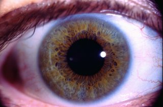
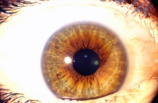
The light brown 'halo' around the pupil indicates an over-acid stomach in this third over-exposure of a dark brown iris. According to Dr. Bernard Jensen (personal communication, 1997) very, very few people have a true over-acid stomach; and those with stomach ulcers typically have an under-acid stomach.

In this particular instance, the over-acid stomach sign was most probably a result of the client drinking in excess of three liters of dilute phosphoric acid (soda pop) per day while he was painting houses in the summer.

The 3-4 concentric circles indicate stress to the nervous system, possibly as a result of the leaching of calcium and magnesium in an effort to buffer the massive quantities of phosphoric acid ingested. (In such instances, it would be recommended that the client have his or her doctor test for an electrolyte imbalance.)

Although there has not been any follow-up since the taking of this picture, it is assumed that the over-acid stomach sign resolved once the client began drinking water rather than soda pop.

Outline for Research Presentation

Iridology Research Presentation

February 17, 2006


Slide 1

The ‘sub-acute inflammation’ sign for an under-acid stomach.

I. Medical implications:

A. The inability to completely digest and assimilate animal proteins, resulting in diminished similation of the B vitamins (responsible for brain and nerve activity), and an increase in the homocysteine level (vs. the ‘cholesterol hypothesis’ as the cause of heart disease)

B. The inability to assimilate minerals:

1. Calcium—muscle wasting, osteoporosis

2. Magnesium—Vitamin B cofactor; hypertension

3. Iron—anemia

4. Chromium—insulin co-factor

5. Zinc—immunity, prostate, thyroid, taste, retina (macular degeneration)

6. Selenium—antioxidant, necessary for formation of glutathione peroxidase (a major liver detoxifier)

C. The inability to protect against ingested viral, bacterial and other pathogens.

1. The acute sign in the Peyer’s Patches area at 2:30 o’clock within the under-acid stomach ring is a graphic confirmation of the research contained in the article “Local Hormone Networks and Intestinal T Cell Homeostasis” published in the March 28, 1997 issue of “Science” magazine.


II. Cognitive implications

1. Magnesium/Vitamin B 12 deficiency depression and/or dementia

2. Attention Deficit Hyperactivity Disorder

3. Alzheimer’s Disease/dementia

a) Namenda (an analogue of amantadine, an anti-viral blocking agent)

b) aluminum toxicity

Slide 2 A true/diet-induced over-acid stomach sign in a brown iris

Slide 3

Over-acid stomach ring in a brown eye.

I. Medical implications:

1. Possible electrolyte imbalance due to mineral leaching—osteoporosis, hypertension

II. Cognitive Implications:

1. Attention Deficit Hyperactivity Disorder

2. Magnesium deficiency-->Vitamin B 12 deficiency-->depression


Chronic to Degenerative Lesion in Prostate

Client presented asymptomatically for a 'free iridology screening'; denied any problems or symptoms relating to the prostate.

Chronic to degenerative lesion observed (at ~7:00 o'clock, .25-.50R) indicating problems with the prostate. Chronic to degenerative lesion also observed at the splenic flexure of the colon (at ~1:00 o'clock, .25-.50 R) (Lesions are much sharper and deeper on original image.) Recommended that he see a doctor for further evaluation.

Dr. returned a diagnosis of a chronic prostatitis which, however, was refractory to multiple anti-biotics.

Client had taken several courses of a variety of anti-biotics over several years for annual or semi-annual URIs (upper respiratory infections) without, however, supplementing with acidophilus, bifidophilus or multi-dophilus cultures. This suggests that the resulting prostatitis was the consequence of the elimination of the beneficial bacteria from the colon.

Inasmuch as the client was experiencing no symptoms, this slide raises the question of whether those who are ultimately diagnosed with prostate cancer do/did not have a long-term undiagnosed chronic prostatitis which weakened the prostate gland, making cancer that much more likely. Thus, clients with these particular signs in their iris would be much more effectively served by the test for prostate specific antigen than those who do not have such signs; which, of course, would help in reducing health care costs.

In addition, Dr. Bernard Jensen referred to this pairing of chronic signs in the prostate and the splenic flexure of the colon as a 'prostate syndrome' because of the number of times he had observed such a pairing.

Dr. Jensen also suggests that assessment of prostate problems on the basis of the iris is much more accurate as well as, shall we say, 'less invasive' than the common test for prostate problems. (Benign prostatic hyperplasia or hypertrophy, however, typically presents as an acute--i.e., inflammation--sign rather than a chronic to degenerative sign.)

In this particular case, it was recommended that the client undergo a series of colonics and other natural modalities and dietary changes to correct the fundamental problem of colon toxicity. Additionally, foods with high levels of zinc (to improve immunity as well as support the prostate) would be recommended--soaked and drained (to deactivate the enzyme inhibitors) pumpkin seeds, for example.

Chlorine and Heavy Metal (?) Toxicity in a Blue (yes, blue) Iris

The darkened area around the periphery of the iris is referred to as the 'scurf rim'; which, in this iris, indicates severe under-activity of the skin and/or an accumulation of toxins. (This is also, typically, the sign seen in the irises of women on the cover of 'glamour' magazines as a result of the over-application of cosmetics which do not allow the skin to 'breathe'.)

Black density at ~8 o'clock and 1/4th the distance from the pupil to the periphery of the iris (8:00, .25R) is in the area of the gall bladder (which, similar to the thyroid, is, according to Dr. Bernard Jensen, an 'iodine' organ) and suggests severe under-activity of the gall bladder and the posssibility of a gall stone (which can be ruled out by an ultrasound ordered by a doctor). Also recommend tests for thyroid function.

Possible under-acid stomach ring (~6:30-8:30, .25R).

Yellow tinge to iris indicates over-acidity to the body (recommend alkalinizing diet); orange and reddish-brown coloration is suggestive of possible heavy metal toxicity. (Recommend intravenous and/or oral chelation; cilantro (?); horsetail and stinging nettle teas (?)

Client reported a severe reaction to being placed on a beta blocker for hypertension; was, for a number of years, a member of a synchronized swimming team, which required long hours of exposure each day to chlorinated swimming pool water. (Recommend ozonated swimming pool water, as is common in Europe.)

Size of pupil is suggestive of adrenal fatigue (vitamin C? astragalus?)

Chronic to Degenerative Sign for an Abdominal Aortic Aneurysm


At the time of open heart surgery (CABGX4), client was diagnosed with a 4.5 cm abdominal aortic aneurysm; the sign for which, in this iris, is a chronic to degenerative lesion which occurs at ~4:30 o'clock, .25-.45 R. Also of note is a chronic sign in the area of the spleen (~4:15 o'clock, .25-.35 R) and a chronic to degenerative lesion (~7:30 o'clock, .25-.45 R) which suggests a possible herniation to the lumbar-sacral disk. (Client was experiencing severe pain down his left leg.)

Also of interest is that the client had taken a statin drug for 24 years.Thus, the insurance company ended up paying a significant amount of money for both the statin prescription as well as the open-heart surgery.

Applying the "Web (Solid)" option of the Microsoft Image Composer to the original image (this image could not be uploaded) demonstrates that the lesions in the iris are both color and depth lesions: the lesions in the abdominal aorta and the lumbar spine are sharpened and deepened; whereas the chronic lesion in the spleen is 'washed out' as being mostly a color rather than a depth lesion.

The yellow density--xanthelasmus--lateral to the edge of the iris is suggestive of hyperlipidemia, which is probably the reason the client was prescribed a statin drug in the first place; despite the lack, however, of the classic iris sign for hardening of the arteries (as seen below)

Classic Iris Sign for Hardening of the Arteries

The white density along, mostly, the medial and superior edge of this iris is the classic iris sign for hardening of the arteries and is typically referred to as a sodium/cholesterol ring, although a lipid panel will not always indicate hyperlipidemia.

Also of interest is a chronic lesion (weakness) in the "peritoneum/abdominal wall" area of the iris (~5:30 o'clock, 0-.20R).

Client experienced a sharp pain while shovelling snow approximately three weeks after this picture was taken. He went to his doctor and was diagnosed with a left inguinal hernia.

Chronic sign at ~4:06-4:18 o'clock, .15-.25R in the spleen area, almost completely surrounded by an acute sign. Clinical significance unknown.

Diagnosis: R/O Inflammatory Breast Cancer


Client presented to her doctor with symptoms involving her right breast and had undergone a series of tests to determine the cause. Her doctors were unable to agree on a definitive diagnosis but had a high 'index of suspicion' for inflammatory breast cancer.

Since the client was not a smoker, the strong blue tinge to the periphery of most of the iris suggests tissue hypoxia and a possible iron deficiency; but the probable cause for such a deficiency cannot be determined by the iris.

The sign of particular interest is what appears to be a chronic to degenerative lesion at 8:30 o'clock, ~.6R; which is not the 'classic' breast cancer sign observed by Dr. Jensen. A similar sign also occurs in the esophagus area at 3:06 o'clock, ~.6R, and in the lung area at 9:36 o'clock and .6R.

Months subsequent to this picture, it was provisionally determined that the client did not have inflammatory breast cancer; a conclusion which demonstrates both the power and the limitations of Iridology.

The existence of chronic to degenerative signs in both the breast area, the esophagus area and the lung area raises the question of whether the client's mother or father was a smoker who had suffered breast, lung and/or esophageal cancer prior to the client's conception. That is, some chronic to degenerative signs in the iris are a result of genetic inheritance and do not necessarily indicate significant problems with the client's physiology.

Thus, Iridology is a science in which 'false positives' tend to outnumber 'false negatives'.

In addition, organs removed under anesthesia retain the sign prior to their removal--an acute sign in the appendix or tonsils after an appendectomy or tonsillectomy, for example--because anesthesia disrupts the direct neurological connection between that organ and the iris.

According to Walter Lang, a German medical researcher, 'autonomic nerve fibers from every tissue in the body connect to the thalamus and hypothalamus; and, from there, through the opthalmic branch of the trigeminal ganglion to the motoneurons of the iris structure.'

("Iridology, the Science and Practice in the Healing Arts, Volumn II", Bernard Jensen, Copyright, 1982, pg. 84)

'Sub-acute Inflammation' (Under-acid stomach) in a Smoker

Although difficult to see in the uploaded image, this is what an under-acid stomach looks like in the iris of a smoker: the light blue-gray 'halo' around the pupil tinged with discoloration.

Client reported digestive problems, especially with proteins, as well as very bad breath.

Blue tinge across the upper edge of the iris indicates hypoxia (diminished oxygenation) to the brain. Hypoxia may result from either decreased circulation, decreased hemoglobin, or decreased 'carrying capacity' of the hemoglobin. Inasmuch as this client does not have the classic sign for hardening of the arteries, and the hemoglobin number is unkown, the hypoxia to the brain is probably the result of smoking--i.e., the level of carboxyhemoglobin--which can be determined by a test ordered by a doctor.

Diagnosis: Breast (sic!) Cancer (Fatal)


Client presented with a very recently diagnosed 'aggressive' breast cancer and breast removal; expired approximately 3-4 months after the taking of this picture.

This is the fourth over-exposure of a dark brown right iris revealing a chronic lesion in the rib area (8:06-8:30 o'clock, .5R) and, perhaps, an acute (inflammation) sign (white area immediately superior to red-brown chronic dot lesion) in the breast area. (Such acute signs also appear in the irises of 13 year old girls, where they signify not inflammation, but the rapid growth of breast tissue; and in the irises of nursing mothers, indicating increased tissue activity.) Possible acute sign and/or faint "lymphatic rosary" through the pleural space (8:54-9:00 o'clock, .6-.8R), suggesting lymphatic involvment and either a clinical or sub-clinical 'pleural friction rub'.) There is no 'classic' sign for breast cancer (Bernard Jensen, 1952); nor is there any clear evidence of significant breast involvement with a degenerative disease process.

Client had been taking birth control pills (with progestin and estradiol--phonetically, estra-DIE-all) for more than 17 years; had complained to her doctor for more than a year of significant pain in her right rib area, but had been unable to convince him to do additional testing other than an X-ray, which was read as 'normal'.